Ear Anatomy – Tiny Pieces with Big Jobs
Ear anatomy is not as complex as you might think. There are only a few anatomical parts that make hearing possible. However, if there is something wrong with any one of those small pieces, you can lose your hearing completely.
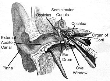
The ear is made up of three parts–the outer ear, middle ear, and inner ear. Each of these small parts in ear anatomy holds a specific job in the process of detecting and interpreting sound.

Outer Ear
The part of the outer ear that you can see on the sides of your head is called the ear flap, or pinna. The pinna is the part of your ear anatomy that collects sounds and protects your middle ear from damage to your ear drum.
Your external auditory canal is just inside your pinna and is also part of your outer ear. Sound travels through this canal.
Your ear drum (tympanic membrane) is what separates your outer ear from your middle ear. Your ear drum is a tightly stretched structure that is located at the end of your external auditory canal.
Middle Ear

Your middle ear is the part of your ear anatomy that is an air-filled cavity. It contains the three tiniest bones in your body. These bones are known as your ossicles.
The names of the three individual bones of your ossicles are: the malleus (the hammer), the incus (the anvil), and the stapes (the stirrup).
Your ossicles stretch across your middle ear. Your malleus is attached to your ear drum, and your stapes is attached to your oval window.
Your oval window separates your middle ear from your inner ear.
Inner Ear
Your inner ear is made up of two organs with different functions.
Your semicircular canals are chambers filled with fluid that do not do anything for your hearing. Instead, they help maintain your body’s balance and equilibrium (they keep you from being labeled as “klutzy”).
Your cochlea is the second of the two organs of your inner ear. This is your body’s microphone. It looks like the shell of a snail, and is divided into three chambers that are filled with fluid.
The central chamber of your cochlea is the part of your ear anatomy that contains your organ of corti. Your organ of corti has between 15,000 and 20,000 nerve cells (that look like tiny hairs) attached to it. These tiny cells are connected to your auditory nerve (your nerve of hearing) which carries signals to your brain.
Hearing
Sound, which is anything from rain, music, and speech to someone’s annoying singing, sends vibrations (sound waves) into the air.
Your pinna (ear flap) collects these sound waves. The sound waves then travel through your external auditory canal and strike your ear drum (like a drum). Your ear drum then vibrates at the exact same frequency of the sound your pinna originally collected.
The vibrations then pass through your malleus, incus, and stapes in your middle ear (which again, vibrate at the same frequency as the sound wave).
The vibrations move to your oval window, then the fluid of your cochlea, and then to the tiny hair-like nerve cells in your cochlea’s central chamber.
These nerve cells each have sensitivity to a specific frequency of sound. The nerve cells that have a natural frequency that matches the sound wave’s frequency will then resonate with that vibration.
The cells then produce an electrical impulse that travels along your auditory nerve to your brain.
Finally, your brain interprets the impulses as sound.
Amazing! Ear anatomy is truly very interesting. These tiny bones and organs complete one of our senses. And, if anything is every wrong with one of them, the entire system is skewed and you lose your hearing.
Hearing Damage
We live in a noisy world. Noise may come from our work or from voluntary exposure to noise, such as noisy motors or loud music at rock concerts, night clubs, discos and from stereos – with or without the use of headphones. Also the increasing use of portable music players are causing hearing damages. The players are capable of delivering high sound levels and the user risks exposing their ears to highly excessive dB levels.
More Information
For more information about this topic, you can visit the All About The Human Ear website.
You can also see the human ear in 3D on this website: Human Ear in 3D.


